
Experimental device yields fresh hope for patients with spinal cord injuries
May 8 (UPI) -- A combined science-medical team at Cambridge University in England has developed a new minimally invasive technique to treat spinal cord injuries with a miniature, wrap-around, electronic implant to comprehensively map nerve signals.
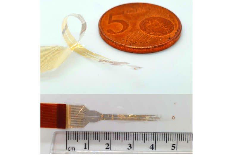
Unlike current high-risk approaches that involve cutting into the spinal cord to fit electrodes and placing implants in the brain, high-resolution implants a few microns thick are wrapped around the entire circumference of the cord to provide a 360-degree picture of nerve activity, the university said in a news release.
Tests in live animal and human cadaver models showed the devices also could stimulate limb movement and bypass injuries in which communication between the brain and spinal cord had been totally lost.
The research, published Wednesday in the U.S. journal Science Advances, could pave the way for treatments without the need for risky brain surgery, and while that is several years away, the team's plan is to use the devices to reliably monitor spinal cord activity during surgery.
"Most technologies for monitoring or stimulating the spinal cord only interact with motor neurons along the back, or dorsal, part of the spinal cord," said Dr. Damiano Barone, of the Department of Clinical Neurosciences, who co-led the research.
"These approaches can only reach between 20% and 30% of the spine, so you're getting an incomplete picture," Barone said.
The spinal cord is notoriously difficult to study, so greater understanding could lead to improved treatments for a range of conditions, including chronic pain, inflammation and hypertension, he said.
With a thickness of just a few millionths of a meter, the Cambridge-developed biocompatible devices are made from advanced photolithography and thin film deposition techniques and require minimal power.
They work by intercepting signals travelling on the nerve fibers of the spinal cord, allowing the signals to be recorded. The thinness of the devices means they can record the signals without causing any damage to the nerves, since they do not penetrate the spinal cord.
"It was a difficult process, because we haven't made spinal implants in this way before, and it wasn't clear that we could safely and successfully place them around the spine," said co-lead George Malliaras, of Cambridge's Department of Engineering.
"But because of recent advances in both engineering and neurosurgery, the planets have aligned and we've made major progress in this important area," he said.
Rat model tests successfully stimulated limb movement by using an adaptation of a routine surgical procedure allowing the devices to be slid under the spinal cord without damaging it, researchers said.
Reaction time was close to human reflexive movement with additional tests using human cadaver models also proving successful, they said.
The researchers said their approach could change how spinal injuries are treated by removing the need for the brain implants required by current techniques.
"If someone has a spinal injury, their brain is fine, but it's the connection that's been interrupted. As a surgeon, you want to go where the problem is, so adding brain surgery on top of spinal surgery just increases the risk to the patient," Barone said.
This vital field of anatomy has long been understudied because the structures are so delicate and complex as to make it near impossible to directly study the whole of the spinal cord in a human.
However, Barone said the monitoring nerve activity during surgery approach showed "enormous potential" for helping patients.
The research was conducted with the help of funding from the Royal College of Surgeons, the Academy of Medical Sciences, Health Education England, the National Institute for Health Research and the Engineering and Physical Sciences Research Council.

